Search the news
Total: 244
article image

TCTAP 2023

P2Y12 Inhibitor or Asprin for Long-term Antiplatelet Management

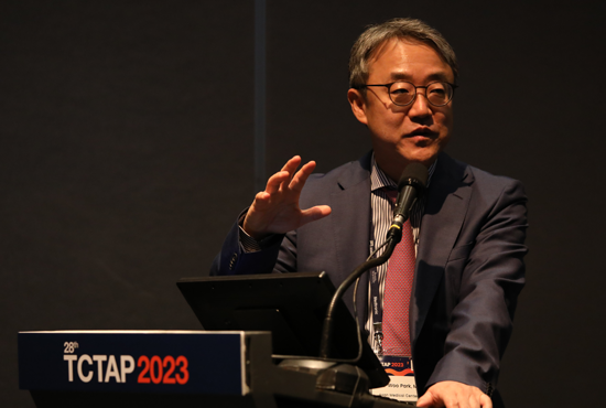
Kyung Woo Park, MD, PhD (Seoul National University Hospital, Korea), introduced the latest evidence on long-term antiplatelet management, including their HOST-EXAM trial at TCTAP 2023 on May 7th. Antiplatelet therapy is the mainstay of treatment to reduce the ischemic risk in patients undergoing percutaneous coronary intervention (PCI). The current guidelines recommend life-long single antiplatelet therapy (usually aspirin as the first-line treatment) following a period of dual antiplatelet therapy (DAPT). However, this recommendation was not based on a large, randomized trial. Therefore, the comparative efficacy and safety of monotherapy with a P2Y12 inhibitor or aspirin after a due duration of DAPT still remains incompletely understood in post-PCI patients. In the HOST-EXAM trial, patients undergoing PCI with a drug-eluting stent who completed 6-18 months of DAPT without any ischemic or major bleeding complications were randomized to monotherapy with clopidogrel or aspirin. The primary endpoint was a composite of all-cause death, non-fatal myocardial infarction (MI), stroke, readmission due to acute coronary syndrome, and bleeding academic research consortium (BARC) type ¡Ã3 bleeding. At 24 months, the use of clopidogrel was associated with a 27% relative reduction in risk of the primary composite endpoint. The benefit of clopidogrel was also observed for the thrombotic composite outcomes as well as any bleeding. ¡°In the HOST-EXAM Extended study, we extended follow-up of these patients up to median 5.8 years after initial randomization,¡± he mentioned (Figure 2). Figure 1. Study flow of the HOST-EXAM Extended study. In the HOST-EXAM Extended study, the composite primary endpoint occurred in 12.8% of the clopidogrel group compared to 16.9% of the aspirin group at a median follow-up of 5.8 years. (HR 0.74; 95% CI 0.63-0.86; p

May 24, 2023 2048

article image

TCTAP 2023

State-of-the-Art Technique for Transcatheter Closure of Paravalvular Leaks

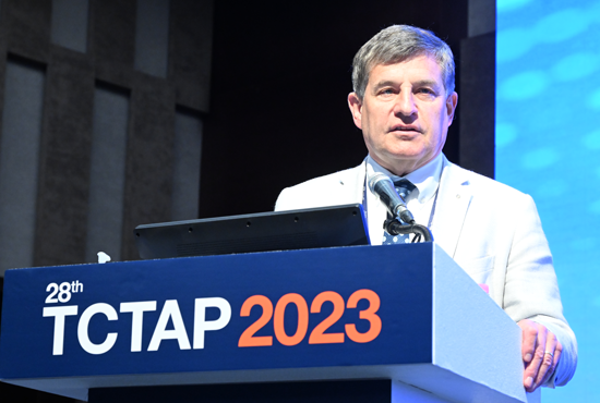
Renowned cardiologist, Wojciech Wojakowski, MD (Medical University of Silesia in Katowice, Poland), sheds light on technical considerations and execution of the transcatheter paravalvular leak (TPVL) procedure during a recent medical conference. Paravalvular leaks (PVLs) are serious complications that can occur after surgical valve replacement or transcatheter aortic valve replacement (AVR). TPVL closure offers a less invasive alternative to surgical re-intervention. The safety and feasibility of TPVL closure have been validated through several registries and a meta-analysis. He shared valuable insights regarding technical considerations and recommendations for the TPVL procedure on May 8th, 2023, at the TCTAP conference. "PVL occurs due to various factors such as the type of prosthesis and endocarditis," he explained. "The overall incidence of PVLs ranges from 5% to 18%, with 74% occurring within the first-year post-operation and 16% appearing later due to suture dehiscence associated with subacute bacterial endocarditis." Indications for TPVL Closure He emphasized that TPVL is contraindicated in the presence of valve rocking/instability or active infective endocarditis. However, the procedure can be performed when heart failure symptoms (NYHA II-IV class) and/or hemolysis are present, along with a PVL jet of Grade 2 or higher observed in color Doppler (color coded Doppler flow mapping) on echocardiography. Additionally, specific parameters related to the valves need to be identified. For mitral PVL, these parameters include systolic flow reversal in the pulmonary vein, increased pulmonary artery pressure, lack of left atrium (LA) size reduction, or progressive LA dilatation after mitral valve replacement (MVR), and high forward transprosthetic flow velocity. In the case of aortic PVL, parameters, such as holodiastolic flow reversal in the descending aorta, lack of left ventricle (LV) size reduction, or progressive LV dilatation after AVR, and high forward transprosthetic flow velocity, should be assessed. Diagnosis of PVL When PVL is suspected, an echocardiography study is crucial to confirm the diagnosis. Three-dimensional transesophageal echocardiography (3D-TEE) allows for a better definition of the PVLs, assessing their number, shape, and location, making it the gold standard for PVL evaluation. "For PVL sizing, the cross-sectional area can be measured in 3D-TEE multiplanar reconstruction, using single-beat acquisition only," he added. TEE plays a crucial role during the TPVL closure procedure, and 3D-TEE is essential for device selection, procedure guidance, and result assessment. Additionally, fusion imaging techniques automatically fusing live 3D-TEE and live X-rays in real-time can provide valuable assistance during procedures such as a transseptal puncture or crossing the PVL with the guide wire. Figure 1. Transcatheter Techniques for TPVL Closure When it comes to aortic PVL closure, the retrograde approach is commonly used for most cases, while the anterograde approach with an apical puncture is rarely employed. He explained the "mother-and-child technique" which involves using a diagnostic catheter within a guiding sheath. After crossing the defect, a hydrophilic guidewire is replaced with a stiffer wire, and the device is finally deployed after advancing the delivery sheath. In the case of mitral PVL closure, the anterograde approach with transseptal puncture is mostly utilized, while the retrograde approach with transaortic or transapical puncture is seldom used, particularly for posterior or medial mitral PVL. He emphasized that the location of the septal puncture depends on the PVL's location and should be guided by TEE. In certain instances, such as septal PVL or unfavorable angles between the transseptal puncture and the defect, using a deflectable catheter like the Occlutech steerable guiding sheath can be highly beneficial. Current Device Selection PVLs vary in size and shape, often appearing crescentic and serpiginous rather than cylindrical, making it challenging to find a single device suitable for all cases. The Amplatzer Vascular Plug (AVP) III is widely used and approved for PVL in Europe. The Occlutech Paravalvular Leakage Devices (PLD) have received a CE-marking for PVL. According to him, AVP III lacks sealing materials on both sides of the disc, allowing for oversized use in irregular or crescentic PVL and multiple PVLs (Figure 2). On the other hand, PLD features sealing materials on both sides of the discs, not permitting oversizing and enabling use in round or oval PVL but limited to single PVL cases. Figure 2. Current device selection for PVLs Post-Procedural Assessment and Follow-Up Multiple registries and a meta-analysis have confirmed the safety and feasibility of TPVL closure. However, there is a risk of residual or recurrent leak and hemolysis, and incomplete closure, despite reducing heart failure symptoms, may lead to a worsening of hemolysis. He highlighted factors influencing hemolysis recurrence after TPVL, including the amount of remaining paravalvular leakage (less than 90% of the cross-sectional area), mitral location, and calcification. Post-procedural imaging with TEE is recommended to assess device position and residual regurgitation, utilizing color Doppler imaging and specific parameters such as pulse wave Doppler of the pulmonary vein for mitral PVL. Future Directions He concluded that transcatheter closure of PVL is the preferred treatment for patients with heart failure or hemolytic anemia compared to surgical re-intervention, offering lower procedural morbidity and mortality. However, to further enhance procedural success and outcomes, there is a need for dedicated devices that provide complete sealing.. Meet the Experts over Breakfast Mitral & Tricuspid Valve Intervention Monday, May 8, 7:00 AM - 8:00 AM Valve & Endovascular Theater, Vista 1, B2 CHECK THE SESSION

May 24, 2023 1895

article image

TCTAP 2023

Radiation Management for Complex Percutaneous Coronary Intervention

According to Gerald Werner, MD (Klinikum Darmstadt GmbH, Germany), percutaneous coronary intervention (PCI) has become a cornerstone in the management of coronary artery disease (CAD). As the complexity of PCI increases, so do the duration of the procedures and the radiation exposure for both patients and medical staff. This increased radiation exposure can have substantial long-term health effects. Therefore, effective radiation management strategies are necessary to ensure the safety and the effectiveness of complex PCI procedures. It is a fundamental principle of radiation physics that radiation cannot be eliminated, only diminished. Therefore, in the context of PCI procedures, our goal is not to eliminate radiation exposure, but rather to manage and reduce it to the lowest levels that are reasonably achievable. However, a significant challenge lies in the fact that operators are often unaware of the ways to optimize their radiation use. Familiarity with X-ray equipment settings, understanding the readings and their implications, and a keen interest in dose optimization are vital components of effective radiation management. A variety of factors influence the radiation dose in PCI procedures, including the complexity of the lesion, the patient's body weight, and the quality of the X-ray equipment. Complex lesions, for example, can cause the operation to be prolonged, resulting in greater radiation exposure. Patients with a higher body mass, on the other hand, may require a higher radiation dose due to the increased demand for tissue penetration. A mix of modern imaging technology and clever procedural approach is required to address these obstacles and decrease radiation exposure. Recent technological advances, such as newer-generation flat-panel detectors, provide high-quality imaging at substantially lower radiation doses. These technical advances, however, must be accompanied by meticulous procedural planning. The optimization of procedure parameters is a critical component of radiation dose reduction. This involves minimizing fluoroscopy duration, employing the lowest radiation dose possible for image acquisition, and adjusting technical features, such as fluoroscopy frame rates to the unique needs of each patient. Furthermore, measures like appropriate shielding, proper collimation, reducing the angle of C-arm, and saving fluoroscopic images instead of taking other Cine images play an important role in further reducing radiation exposure. There is no doubt that managing radiation exposure in sophisticated PCI procedures is difficult. A solid understanding of radiation physics, on the other hand, can go a long way toward ensuring that radiation is successfully attenuated. Furthermore, ongoing education and training are required to effectively implement these tactics. By implementing these steps, we can ensure that complicated PCI remains a safe and effective treatment choice for CAD patients. Hot Topics Complex PCI - Calcification, Very Long Lesion Monday, May 8, 2:50 PM - 4:00 PM Presentation Theater 1, Vista 3, B2 CHECK THE SESSION

May 18, 2023 2282

article image

TCTAP 2023

Great Debate on MVD 2023: Medical Therapy is Enough vs. Surgery is Still Standard

Sripal Bangalore, MD (New York University Grossman School of Medicine, USA) and David Paul Taggart, MD (University of Oxford, UK), had a heated debate regarding the treatment of multi-vessel disease (MVD), with Dr. Bangalore advocating for medical treatment and Dr. Taggart favoring surgery. Dr. Bangalore began the debate by acknowledging that coronary artery bypass grafting (CABG) can improve survival rates. He also argued that the results of the ISCHEMIA trial, which studied the effectiveness of different treatments for stable ischemic heart disease (IHD), do not necessarily apply to CABG. Additionally, he asserted that neither medical therapy nor percutaneous coronary intervention (PCI) reduces the risk of myocardial infarction (MI) in patients with MVD. Thus, he brought up the debate topic of whether CABG improves overall survival in patients with MVD and preserved left ventricular systolic function. Dr. Bangalore presented a recent meta-analysis showing routine revascularization was not associated with improved survival compared with medical treatment in patients with stable IHD (Figure 1). In addition, he referenced the BARI-2D CABG Stratum and MASS II 10 years follow-up, which did not show the mortality benefit of CABG over guideline-directed medical therapy (GDMT). He also stated that the ISCHEMIA trial included a larger number of patients who underwent CABG than other clinical trials that have compared revascularization with medical treatment, but it did not conclude that revascularization is superior to medical treatment. Unlike PCI or CABG, advanced medical therapy provides a systemic approach to the treatment of MVD. This approach involves the use of various medications, including beta blockers, ACE inhibitors or ARBs, statins, PCSK9 inhibitors, DOACs, antiplatelet medications, and SGLT2 inhibitors. These medications have been shown to provide survival benefits and improve cardiovascular clinical outcomes in patients with MVD. In some cases, advanced medical therapy may be an alternative to revascularization procedures such as PCI or CABG, especially in patients who are not candidates for these procedures due to various reasons such as comorbidities or personal preferences. He concluded that medical therapy, with or without PCI, is sufficient for most patients with MVD with CABG reserved for those with complex coronary disease. Figure 1. The Fallacy of CABG benefits David Paul Taggart University of Oxford, United Kingdom Dr. Taggart still maintains the view that CABG is still the standard treatment approach for patients with MVD. First of all, he emphasized three cautions and key rules for interpreting trials and data of PCI vs. CABG; 1) the trial patients may not represent the typical patient population encountered in routine clinical practice (Figure 2). In other words, the patients in clinical trials may have less severe coronary artery disease (CAD) than patients encountered in routine practice. Therefore, the benefits of CABG observed in clinical trials may be underestimated in routine practice where most patients have more severe CAD. 2) the duration of follow-up is critical when interpreting data from trials comparing PCI and CABG. He noted that a minimum of 5 years of follow-up is necessary, with an ideal follow-up period of 10 years, as seen in trials such as ART, SYNTAXES, and STITCH. The longer the follow-up period, the greater and more accelerated the benefit of CABG becomes. 3) the use of CABG with GDMT has been shown to have even greater benefits over PCI. Therefore, it is important to ensure that patients undergoing CABG receive optimal medical therapy to maximize the benefits of the procedure. Dr. Taggart presented various clinical trials, including SYNTAX, FREEDOM, FAME III, and STITCH trials, as well as observational studies, to demonstrate that CABG provides superior mortality and cardiovascular outcomes compared to PCI or medical treatment alone. He stressed the effectiveness of CABG by citing the 2018 guidelines on myocardial revascularization which classified CABG as Class I and highlighted that CABG could be more beneficial with more arterial grafts and optimal medical treatment. Dr. Taggart also gave three reasons for the persisting survival benefit of CABG over PCI. Firstly, anatomically, atheroma is mainly located in the proximal coronary arteries, which makes CABG more effective because it bypasses the diseased segment. Secondly, the internal mammary artery is known to elute nitric oxide (NO) into the coronary circulation, which has a beneficial effect on the endothelium and reduces the risk of further disease. On the other hand, drug-eluting stents used in PCI can impair reendothelialization and downstream endothelial function, which creates a pro-thrombotic environment and may increase the risk of further cardiovascular events. Lastly, PCI can sometimes result in incomplete revascularization, meaning that not all blocked or narrowed vessels are treated during the procedure. In contrast, CABG aims to provide complete revascularization by bypassing all significant blockages in the coronary arteries. He concluded that CABG is considered the standard, but PCI or medical treatment may be considered in cases of multiple comorbidities, frailty, or reduced life expectancy. Figure 2. Three cautions and key rules for interpreting trials and data Hot Topics Left Main & Multi-Vessel (Concept Changes After ISCHEMIA) Sunday, May 7, 3:54 PM - 4:10 PM Presentation Theater 1, Vista 3, B2 CHECK THE SESSION

May 18, 2023 2175

article image

TCTAP 2023

The 20th Anniversary of TAVR Ceremony - From Concept to Human Application

As the recipient of the 13th TCTAP ¡°Master of the Masters¡± Award, Alain G. Cribier, MD (Hospital Charles Nicolle, University of Rouen, France), delivered a lecture entitled ¡°Celebrating 20 Years of TAVR: My Love Story for TAVR.¡± The history of interventional medicine is short with an explosion of innovative treatments emerging at the end of the 20th century and dramatically expanding in the 21st century. For the younger generation of interventional specialists, with the armamentarium of these strategies at their fingertips, it is intellectually impossible to imagine what it was like treating cardiovascular disease less than 50 years ago. For those of us who were witnesses to that period and the developments that followed, it was another world. The treatment of aortic stenosis (AS) is the most prevalent acquired valvular disease, and the first successful implantation of an aortic stented valve in a human, performed on April 16th 2002, is celebrated the 20th anniversary. But before the development of transcatheter aortic valve implantation (TAVI), non-coronary heart diseases, along with their dedicated interventional techniques, saw rapid growth from the transcatheter treatment of congenital pulmonic stenosis in 1979 and aortic valvular stenosis (AVS) in 1983, mitral valvuloplasty in 1984, and balloon aortic valvuloplasty (BAV) in 1985. In September 1985, faced with a 72-year-old female patient with calcified AS, recurrent syncope, and imminent death, the team at Rouen University Hospital adapted an existing technology from the field of congenital valve disease. Under local anaesthesia, a pulmonary balloon was employed to dilate the patient¡¯s aortic valve using a percutaneous transcatheter femoral approach. This first-in-human BAV led to an immediate haemodynamic and clinical improvement, followed by relief of symptoms and a return to normal life for the patient. However, after several years it became evident that balloon dilatation was not a long-lasting solution due to early valvular restenosis in 80% of patients at one-year follow-up. The merciless criticism to the failure of BAV reinforced a resolution to find a viable solution to deal with the issue of aortic valve re-stenosis. Remarkably, this technique knew re-birth with the onset of TAVI since it is used in daily practice to predilate the native valve or post-dilate the prosthesis. The concept of implanting a dedicated stented valve for calcified AS was born out of this challenge. All BAV balloons could fully expand the calcified aortic valve despite the calcification. The idea that emerged was to maintain the initial positive results of the expansion by implanting a stent with a valve inside using the diseased native calcified valve as an anchor. This concept posed other issues besides the nature of the diseased native valve itself. Critical questions arose concerning the immediate proximity of essential anatomical structures: above, the coronary ostia and below, the mitral valve insertion, and the His bundle at the upper part of the interventricular septum. In Rouen, a landmark intensive, in-depth research with autopsies validated the concept of valvular stenting in AS. With my colleague, Helene Eltchaninoff, we did validate the correct dimensions of the stented valve to ensure there would not be damage to the surrounding structure, a key step to guarantee the feasibility and safety of the concept. The stents were strongly anchored within the calcified valve structures, thus decreasing the risk of secondary device embolization. In 2002, the first-in-man implantation of this revolutionary device could be performed as a last resort option in a 57-year-old male patient, with severe AS on a very calcified bicuspid valve. He was dying, in cardiogenic shock, with a 12% left ventricular ejection fraction. He had severe comorbidities, including lung cancer, acute ischemia of the leg by occlusion of an aortofemoral bypass, and a floating thrombus into the left ventricle. For these reasons, he had been turned down for surgical valve replacement despite his age. Against all odds, we could successfully perform this first TAVI case using an unplanned highly challenging transeptal approach since the femoral arteries were not usable. This was followed by a breath-taking clinical improvement. The case reported in Circulation hit the headlines. The technique then expanded first in our center with 38 patients treated on a compassionate basis, then in a few centers in Europe and the USA. In 2005, a total number of 100 patients had received TAVI. The turning point was the acquisition of our start-up by Edwards Lifesciences in 2004, leading to an evolution in the technology that continues to this day. A valve size of 26 mm was added, and a steerable delivery system was created to allow an easier and safer transfemoral approach. This approach was pioneered by John Webb in Canada. In 2005, there was the launch of a concurrent TAVI system, the self-expanding CoreValve later acquired by Medtronic, which played an incontestable role in the fabulous expansion of TAVI worldwide. Rapidly many technological improvements were made year after year, with the development of new TAVI systems, new approaches, lower profile devices, and preventive solutions against paravalvular leaks. Consequently, the simpler and safer transfemoral approach progressively turned into the standard strategy for TAVI, used in more than 90% of patients, leading to a ¡®democratisation¡±¡¯ of the procedure and contributing to the burst of implantations worldwide (Figure 1). Figure 1. Developing TAVI, a long bulky road In parallel with the advanced models of TAVI systems, a careful process of staged evidence-based medicine led to the proof that a treatment, originally limited to ¡®compassionate¡¯ use in high surgical risk patients, could be enlarged by the American and European guidelines to include patients at intermediate risk and finally, in 2019, to low surgical risk patients as well. The acute and long-lasting non-inferiority or superiority of TAVI over surgery was consistently demonstrated on many main or secondary endpoints, more particularly death and stroke rates. The number of patients who benefited from TAVI is now reaching two million, in over 80 countries. In numerous countries, the number of TAVI procedures now exceeds the number of surgical valve replacements (Figure 2). How can be seen the future of TAVI? A continuous expansion of the procedure can be predicted, in the range of 10% per year for several reasons: 1) The aging population, whereas aortic stenosis is typically a disease of the elderly, earlier detection of the disease by regular practitioners. 2) The continuous improvement in technology which has notably increased the safety of the procedure. 3) A growing experience of the individual operators leading to an increase in the number of specialized centers practicing TAVI. When combined, experience and accessibility have made TAVI an easy procedural choice where in the past cardiac surgery would have been the only option. 4) The continued expansion of indications to low-risk younger patients provided that the long-term durability of the implanted valves will be fully demonstrated. This has encouraged research on other types of valves than those made of the pericardium, less prone to deterioration. 5) The continued and future expansion of TAVI to other indications, such as valve-in-valve procedures (such as treating future TAVI dysfunction) or the treatment of moderate AS in patients with heart failure or asymptomatic patients with severe AS. 6) Also, importantly, the increase in TAVI procedures and the vital competition between devices should lead to an overall decrease in the cost of TAVI which is of particular interest for lower-income and developing countries. 7) And of course, the outstanding combination of excellent clinical results and the simplicity of the procedure for the patients themselves: no sternotomy, the use of local anaesthesia, a short hospital stay, and, most importantly, a rapid return to normal life without the need for rehabilitation. Figure 2. Current position of TAVI This steady expansion is an excellent example of successful translational research, and moving findings from concept to bench, bench to bedside, feasibility trials to larger clinical registries, and evidence-based trials to everyday practice. The constructive dialogue that continues between multidisciplinary physicians and engineers has been central to the evolution of TAVI. However, it is of interest that TAVI, unlike PCI, began with the most untreatable of patients and only now is being used in lower-risk groups (Figure 3). In the advances in the treatment for valve disease TAVI played - and continues to play - a seminal role. Looking back over the last 20 years since that first TAVI, we are astonished by the impact this procedure has had. TAVI crystallized the study of structural heart disease, inspiring a younger generation of interventionalists and scientists. It remains an innovative and disruptive technology influencing many of the ways that medicine is practiced today. In cardiology alone, it paved the way for percutaneous transcatheter treatment of the mitral and tricuspid valves, has led to far-reaching developments in cardiac imaging, and has been instrumental in bringing together specialists in the Heart Team concept. After 20 years, the future stories of TAVI are still being written. Figure 3. Predictable future of TAVI Hot Topics Game of Thrones, TAVR vs. SAVR Sunday, May 7, 4:10 PM - 5:30 PM Valve & Endovascular Theater, Vista 1, B2 CHECK THE SESSION

May 06, 2023 2614

article image

TCTAP 2023

10-Year Extended Follow-up of BEST Trial

Jung-Min Ahn, MD, PhD Asan Medical Center, Korea Clinical trials in the era of drug-eluting stents (DES) have shown that percutaneous coronary intervention (PCI) including SYNTAX (Synergy Between Percutaneous Coronary Intervention With Taxus and Cardiac Surgery) and FREEDOM (Future Revascularization Evaluation in Patients with Diabetes Mellitus: Optimal Management of Multivessel Disease) has a higher risk of repeat revascularization, spontaneous myocardial infarction, and mortality in multivessel coronary artery disease (MVCAD) patients. Previous trials using first-generation DES and little use of intracoronary imaging have limited applicability in contemporary practice. However, the new-generation DES have a lower incidence of cardiac events and mortality than first-generation stents, and intracoronary imaging can optimize the PCI results. The BEST (Randomized Comparison of Coronary Artery Bypass Surgery and Everolimus-Eluting Stent Implantation in the Treatment of Patients With Multivessel Coronary Artery Disease) trial, which randomly assigned MVCAD patients to undergo either PCI with everolimus-eluting stents or coronary artery bypass grafting (CABG), demonstrated that CABG significantly reduced the composite rate of death, myocardial infarction (MI), or target vessel revascularization. Nevertheless, there was no significant difference in mortality between the two groups. The present study aims to evaluate longer-term outcomes between the two treatments in MVCAD patients who were followed up for up to 13.7 years. The BEST trial was a prospective, open-label, randomized trial comparing PCI with everolimus-eluting stents and CABG in MVCAD patients and was conducted at 27 international heart centers. The primary endpoint was the incidence of major adverse cardiac events (MACE), such as death from any cause, MI, or target vessel revascularization. The major secondary endpoints are a safety composite of death, MI or stroke, and a composite of death, MI, stroke, or any repeat revascularization. A total of 880 MVCAD patients were randomized, with 438 receiving PCI with everolimus-eluting stents and 442 undergoing CABG. The median length of follow-up after randomization was 11.8 years (interquartile range, 10.6-12.5 years; the maximum follow-up, 13.7 years) without significant difference between groups. The mean age of the patients was 64.5¡¾9.4 years; 41% had diabetes; and 77% had 3-vessel coronary artery disease. The baseline demographics and clinical, angiographic characteristics of the patients were well-matched between the two groups. The SYNTAX score was 24.4¡¾7.8 and complete revascularization was achieved in 50.9% and 71.5% of the patients in the PCI group and the CABG group, respectively. The primary endpoint occurred in 151 patients (34.5%) in the PCI group and 134 patients (30.3%) in the CABG group (hazard ratio [HR], 1.18; 95% confidence interval [CI], 0.88-1.56; P=0.26) (Figure 1A). There was no significant between-group difference in the secondary safety composite endpoint of death, stroke, or MI (28.8% and 27.1%; HR, 1.07; 95% CI, 0.75-1.53; P=0.70) and death from any cause (20.5% and 19.9%; HR, 1.04; 95% CI, 0.65-1.67; P=0.86) (Figure 1B and 1C). However, the incidence of spontaneous MI (7.1% and 3.8%; HR, 1.86; 95% CI, 1.06-3.27; P=0.03) and repeat revascularization (22.6% and 12.7%; HR, 1.92; 95% CI, 1.58-2.32; P

May 06, 2023 1986

article image

TCTAP 2023

Routine Functional Testing or Standard Care in High-Risk Patients after PCI

Lack of Evidence for Routine Functional Testing after PCI Duk-Woo Park, MD, PhD Asan Medical Center, Korea The effectiveness of regular stress functional testing after percutaneous coronary intervention (PCI) in improving clinical outcomes is uncertain. The 2019 European guidelines recommend non-invasive stress testing 6 months after high-risk PCI and coronary angiography 3-12 months later, and non-invasive stress testing 1 year after general PCI. However, the recommendations are weak (Class IIB recommendation, Level of evidence C), relying on expert opinions, and the 2021 ACC/AHA guidelines do not mention them. POST-PCI Trial: Routine Functional Testing vs. Standard Care after High-Risk PCI From November 2017 to September 2019, the POST-PCI investigators randomly assigned 1,706 patients with high-risk anatomical or clinical characteristics who had undergone PCI to a follow-up strategy of routine functional testing (nuclear stress testing, exercise electrocardiography, or stress echocardiography) at 1 year after PCI (849 patients), or to standard care alone (857 patients). The primary outcome was a composite of death from any cause, myocardial infarction (MI), or hospitalization for unstable angina at 2 years. The mean age of the patients was 64.7 years, 21.0% had left main disease, 43.5% had bifurcation disease, 69.8% had multivessel disease, 70.1% had diffuse long lesions, 38.7% had diabetes, and 96.4% had been treated with drug-eluting stents. At 2 years, a primary outcome event had occurred in 46 of 849 patients (Kaplan-Meier estimate, 5.5%) in the functional-testing group and in 51 of 857 (Kaplan-Meier estimate, 6.0%) in the standard-care group (hazard ratio [HR], 0.90; 95% confidence interval [CI], 0.61-1.35; P=0.62) (Figure 1). There were no between-group differences with respect to the components of the primary outcome. At 2 years, 12.3% of the patients in the functional-testing group and 9.3% in the standard-care group had undergone invasive coronary angiography (difference, 2.99 percentage points; 95% CI, -0.01-5.99), and 8.1% and 5.8% of patients, respectively, had undergone repeat revascularization (difference, 2.23 percentage points; 95% CI, -0.22-4.68). Figure 1. Time-to-event curves for the primary composite outcome and the components of the primary composite outcome Further, a landmark analysis performed between 1 and 2 years showed a more than twofold higher incidence of coronary angiography (8.2% vs. 3.3%; HR, 2.57; 95% CI, 1.62-4.09) and revascularization (5.8% vs. 2.4%; HR, 2.44; 95% CI, 1.43-4.16) in the functional-testing group compared with the standard-care group. Yet, there were still no meaningful between group-differences in the rate of death or MI (Figure 2). Figure 2. Landmark analysis for outcomes Despite more testing and repeat procedures based on the regular functional testing results, hard clinical endpoints such as death or MI were not reduced. The result was consistent with the recent ISCHEMIA study, which showed that an initial invasive strategy, as compared with an initial conservative strategy, did not reduce mortality or ischemic events among stable patients with myocardial ischemia. The POST-PCI trial results also showed that more aggressive and invasive testing and treatment did not provide additional clinical benefits over a conservative strategy based on guideline-directed medical therapy. In this multicenter, pragmatic, randomized trial of high-risk patients who had undergone PCI, routine functional testing did not result in a lower risk of ischemic cardiovascular events or death from any cause at 2 years. Therefore, routine surveillance stress testing for post-PCI patients is not recommended unless they exhibit clinical signs or symptoms of stent failure. Clinical Science Late-Breaking Clinical Trials 2023 in Asia-Pacific Monday, May 08, 9:30 AM - 10:50 AM Presentation Theater 1, Vista 3, B2 CHECK THE SESSION

May 06, 2023 1949

article image

TCTAP 2023

PREVENT Trial

Seung-Jung Park, MD, PhD Asan Medical Center, Korea Acute coronary syndrome is commonly caused by the rupture of vulnerable plaque, which often has a mild angiographic appearance. Although systemic pharmacologic management is considered a standard therapy for stabilizing vulnerable plaque, the role of the local treatment to prevent plaque rupture has not yet been determined and has only been tested in small trials. The PREVENT trial is designed to compare the effectiveness of preventive percutaneous coronary intervention (PCI) plus optimal medical therapy (OMT) to OMT alone in patients with functionally insignificant, high-risk vulnerable plaques. The PREVENT trial is a multinational, multicenter, prospective, open-label, active-treatment-controlled randomized clinical trial (Figure 1). Patients with at least one angiographically significant stenosis (diameter stenosis >50%) without functional significance (fractional flow reserve [FFR] >0.80) and vulnerable plaque characteristics in intracoronary imaging are eligible for enrollment. Target lesions should have at least two of four intracoronary imaging criteria for vulnerable plaque; (1) minimal lumen area (MLA) 70%; (3) maximal lipid core burden index (LCBI) in a 4 mm segment >315 by near-infrared spectroscopy (NIRS); and (4) thin cap fibroatheroma (TCFA) as determined by optical coherent tomography (OCT) or virtual histology (VH). Enrolled patients were randomly assigned in a 1:1 ratio to either a preventive PCI on vulnerable plaque using bioabsorbable vascular scaffolds or metallic everolimus-eluting stents plus OMT or OMT alone. The primary endpoint is a target-vessel failure, defined as a composite of death from cardiac causes, target-vessel myocardial infarction, ischemic-driven target-vessel revascularization, and hospitalization for unstable or progressive angina at 2 years. Figure 1. The study design of the PREVENT trial. Enrollment of a total of 1,608 patients has been completed. Follow-up of the last enrolled patient will be completed in September 2023 and primary results will be available by early 2024. The PREVENT trial is the first, a large-scale randomized trial, with adequate power for clinical outcomes to evaluate the effect of preventive PCI on non-flow-limiting vulnerable plaque with high-risk features. The results of this trial will provide compelling evidence to determine whether preventive PCI of focal vulnerable plaques on top of OMT improves patient outcomes compared to OMT alone. Clinical Science Future Perspective on Ongoing Trials from AMC Monday, May 08, 11:00 AM - 12:20 AM Presentation Theater 1, Vista 3, B2 CHECK THE SESSION

May 06, 2023 1977

article image

TCTAP 2023

OCTIVUS Trial

Do-Yoon Kang, MD, PhD Asan Medical Center, Korea The clinical value of intracoronary imaging for percutaneous coronary intervention (PCI) guidance is widely acknowledged. Although optical coherence tomography (OCT) and intravascular ultrasound (IVUS) are the most commonly used intravascular imaging methods for guiding and optimizing PCI in daily clinical practice, there are limited data on head-to-head comparison between OCT-guided and IVUS-guided PCI concerning clinical endpoints. Prior studies (ILUMIEN-3, OPINION, etc.) which compared the two imaging modalities might have been hampered by the use of surrogate imaging endpoints, limited patient numbers, and strict inclusion criteria including relatively simple lesions. Future clinical trials should focus on a large, unselected patient population that is more representative of routine clinical practice. The OCTIVUS trial is an investigator-initiated, multicenter, open-label, pragmatic randomized controlled trial comparing the efficacy and safety of OCT-guided versus IVUS-guided PCI strategies in an all-comers population with minimal exclusion criteria (Figure 1). Due to the lack of stringent exclusion criteria, a large proportion of patients with acute coronary syndrome, multivessel disease, or complex lesions, such as left main, bifurcation, long, or restenotic lesions, the patients who represented those undergoing PCI in contemporary clinical practice, could be enrolled in this trial. PCI optimization criteria are predefined using a common algorithm for online OCT or IVUS. The primary endpoint was target-vessel failure (cardiac death, target-vessel myocardial infarction, or ischemia-driven target-vessel revascularization) at 1 year, which was tested for both noninferiority and superiority. Figure 1. The study design of the OCTIVUS trial. From April 2018 through January 2022, a total of 2,000 patients were enrolled. This year, the primary results will be available. It is anticipated to provide valuable clinical evidence regarding the comparative efficacy and safety of OCT-guided versus IVUS-guided PCI strategies in a broad population of patients undergoing PCI. Clinical Science Future Perspective on Ongoing Trials from AMC Monday, May 08, 11:00 AM - 12:20 AM Presentation Theater 1, Vista 3, B2 CHECK THE SESSION

May 06, 2023 2145

article image

TCTAP 2023

30 Years History of FFR: From the Concept to Current Status - Legend's Perspective

Nico Pijls, MD Catharina Hospital, Netherlands In the early days of coronary intervention, some pioneers in coronary intervention recognized the value of coronary artery pressure measurement. However, reliable techniques and devices to measure coronary pressure did not accurately exist at that time. With the development of the true pressure wire and hyperemic method, fractional flow reserve (FFR) was introduced in the 1990s. FFR is a fraction of mean distal coronary pressure (Pd) to mean aortic pressure (Pa) at maximal coronary hyperemia. Today, FFR is used widely in clinical practice. Furthermore, many non-hyperemic methods to measure coronary pressure also have been developed to avoid the inconvenience that occurs during hyperemic stimulation. However, there are clear distinctions between FFR and non-hyperemic indices. FFR is linearly related to maximum achievable blood flow and this linearity is only present under maximum hyperemic circumstances. Clinically, leaving out hyperemia can miss significant stenosis, especially in younger patients with proximal lesions in large coronary arteries. FFR has a sound scientific basis and experimental validation, is accurate and reproducible with an unequivocal normal value and a clear-cut-off with a narrow gray zone, and is easy to perform. FFR is the link amid stenosis severity, maximal blood flow, perfusion territory, and myocardial ischemia. FFR can assess the severity of many complex lesions and functional improvement after percutaneous coronary intervention (PCI) (Figure 1). Figure 1. FFR calculated by ratio of mean Pd to mean Pa Numerous clinical trials have investigated the role of FFR-guided PCI in comparison with angiography alone, medical treatment, or coronary artery bypass grafting (CABG). For instance, the FAME trial showed lower stent use and lower major adverse cardiac events (MACE) in the FFR-guided PCI group compared to the angio-guided PCI group. The FAME-2 trial showed better clinical outcomes in the FFR-guided PCI group than in the medical therapy group. The FAME-3 trial demonstrated that FFR-guided PCI using current-generation drug-eluting stents (DES) did not meet the criteria for noninferiority compared with CABG among patients with angiographic three-vessel disease. In conclusion, anatomy alone is insufficient to understand the physiologic significance of coronary artery disease. FFR provides superior insight into coronary pathophysiology and greatly improves the correct diagnosis of coronary artery disease. There is incontrovertible evidence for improved outcomes of coronary disease and revascularization by the systematic use of FFR. Opening of TCTAP 2023 & Key Note Lectures Sunday, May 07, 9:30 AM - 10:16 AM Main Arena, Walker Hall, Level 1 CHECK THE SESSION

May 06, 2023 2218

Good People, Good Memories, Good Life!