COMPLEX PCI 2024
Left Main and Multi-vessel Disease: Updated Treatment Concept
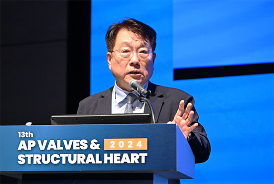
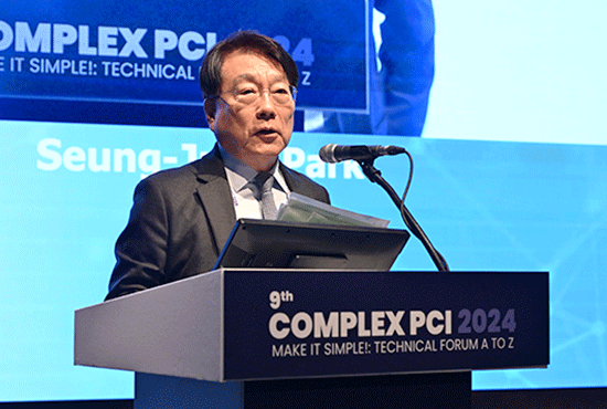
‘Left Main and Multi-vessel Disease: Updated Treatment Concept’ Seung-Jung Park, MD, PhD (Asan Medical Center, Korea), stresses that left main (LM) disease is no longer surgical disease. Until now, LM disease was considered to require surgical revascularization. However, recent clinical trials such as SYNTAX, PRECOMBAT, EXCEL, NOBLE and meta-analysis have led to changes in the recent ESC and ACC/AHA guidelines for LM disease revascularization. 2024 ESC guidelines for revascularization of LM disease categorize LM disease and LM disease with multivessel disease into categories based on SYNTAX score: low complexity is classified as Ia, intermediate as IIa, and for LM disease with multivessel disease, PCI is indicated as IIb if surgical risk is high. 2021 ACC/AHA guidelines classified LM disease revascularization as IIa for low anatomic complexity. However, the global guidelines do not take into account the operator's experience and technical considerations. For example, the left main artery is a large vessel with a proximal lesion and a short lesion length, making it suitable for PCI, and contemporary PCI is physiology- and image-guided PCI, so the global guidelines should reflect this, he pointed. Current Asan Medical Center practice for Left Main Disease There have also been changes in the ESC guidelines for multivessel disease. 2024 ESC multivessel disease revascularization guideline categorize multivessel disease with diabetes and without diabetes. Even in patients with diabetes, PCI is IIa if the surgical risk is high. And in the case of multivessel disease without diabetes, it is almost always Ia. And in the multivessel disease guideline, PCI is no longer classified as III. Current Asan Medical Center practice for Left Main Disease PCI Favour; All ischemic lesions, favourable anatomy for PCI (RVD > 2.5mm, and/or Lesion length < 50mm) CABG Favour; Low EF (< 40%), Diabetic, 3 vessel disease, unfavourable anatomy for PCI Majority of multi-vessel disease, 1 or 2 major vessel PCI with optical medical therapy would be enough. He concluded his lecture by saying that randomized studies are needed to confirm the results because it is not known which outcome is better, contemporary PCI or CABG, in patients with multivessel disease with ischemic cardiomyopathy and multivessel disease with diabetes. Live Case 1: Left Main & Multi-vessel Diseases Friday, November 29, 9:00 AM ~ 10:50 AM Main Arena Check The Session
December 20, 2024 3018