TCTAP 2023
My 30 Years Story - Caring for Patients with Symptomatic Vascular Disease
John Robert Laird, Jr. MD (Adventist Heart & Vascular Institute, USA), shared his journey as a cardiologist in search of surgical strategies for symptomatic vascular disease.
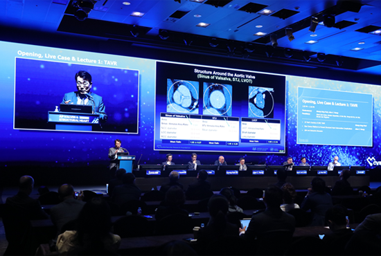
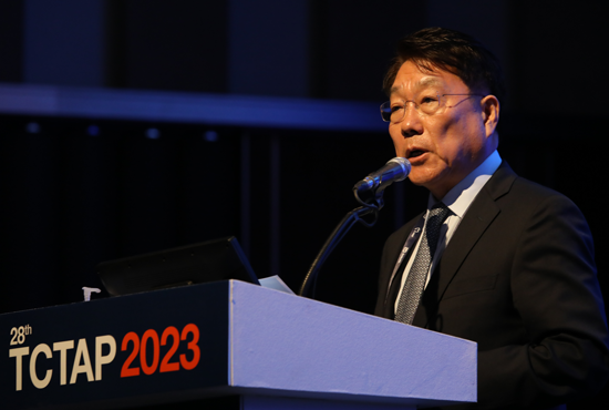
“When reflecting on my past 30-plus years of peripheral intervention as a cardiologist, I couldn't help but think about the people that helped me along the way including my early mentors Dr. Christopher White, Stephen Ramee, and Tyrone Collins at Walter Reed Army Medical Center who were mentors during my cardiology fellowship and first introduced me to peripheral intervention, and then the group at the Washington Hospital Center and Cardiovascular Research Foundation where my colleagues and mentors in my early years as a practicing Interventional cardiologist…(Figure 1).”
Figure 1. Group members of the Cardiovascular Research Foundation in the early years
During his early career, from 1987 to 1990, only balloon angioplasty was available. There were no FDA-approved stents, other devices, or as well as dual antiplatelet therapy. At that time, technical success rates were only 80 to 85% with balloon angioplasty. Problems with acute dissection and elastic recoil made acute vessel closure in 3 to 8% of cases, with significant consequences that were a need for emergency backup bypass surgery were 2 to 5% of cases. Restenosis rates even with successful angioplasty initially were 30 to 50% with balloon angioplasty, so those good old days were not so good. When it comes to iliac artery angioplasty, the results revealed around 90% in 1-month clinical success, but a significant drop off in patency over time-related to restenosis and to disease progression in the iliac arteries. Then, there was a lot of enthusiasm about the new devices that were introduced in the early 1990s including the atherectomy devices and stents. When the stent era began in the 1990s a time frame, the original Palmas 308 iliac balloon stent was presented as 30 mm in length, expandable tubular slotted stent, and could be expanded from 8 to 12 mm in diameter (Figure 2).
Figure 2. Equipment needed for the iliac artery stenting
Over the ensuing years, continued improvements were observed with our stents, both, balloon-expandable and self-expanding stents, and covered stents for iliac artery use and with a good technique such as using intravascular ultrasound guidance. The long-term results of the iliac artery stenting are remarkably good with 15-year primary and secondary patency rates that were over 90%, and we could treat very complex cases such as chronic occlusions (CTO) of both two iliac arteries. As a consequence of excellent angiographic results and good clinical outcomes, we've gone from an era of the bifemoral bypass which was a very commonly performed procedure to an era where it's a very rarely needed procedure because of the excellent results we achieved with treatments in the iliac artery endovascular treatments in the iliac arteries.
Superficial femoral artery (SFA) disease has been much more challenged due to the nature of the disease with diffuse, long, and heavy calcified lesions associated with disease of the infra-popliteal runoff vessels and then the complex mechanical forces that complicate the placement of endovascular prostheses. Because of these challenges, we've seen a host of different technologies, multiple different balloons such as hot balloons, cold balloons, cutting balloons, scoring balloons, and more recently drug-eluting balloons (DEB). There are a whole host of CTO devices that have been developed laser as well as atherectomy devices and a variety of different stents, including, standard laser-cut nitinol stents, covered stents, a woven nitinol stent, and most recently paclitaxel-eluting stents for the SFA and popliteal arteries.
Probably the biggest advance that we've undergone procedures in the SFA has been the advent of the DEB. This was the initial paper from 2004 by Dr. Sheller and Dr. Speck who were able to demonstrate that if you took paclitaxel and mixed it with iodinated contrast, you could then coat a balloon and allow the paclitaxel to come off the balloon and effectively deliver the drug into the vessel wall to inhibit subsequent new intimal proliferation. He was one of the co-principal investigators for the impact SFA drug-coated balloon trial where we saw three years by duplex ultrasound dramatically better results with the Impact-Admiral drug-coated balloon compared to standard balloon angioplasty. We learned better angioplasty techniques because DEB uses long balloon inflations and appropriately sized balloons to avoid geographic mismatch. We could see outstanding results even in complex morphologies in the SFA and popliteal arteries. And we now have data out to five years from the randomized trial as well as the global registry showing sustained benefit with Impact-Admiral drug-coated balloon for complex lesions in the SFA.
Regarding better devices for calcium, atherectomy devices can be used in a very complex calcified lesion followed by DEB angioplasty. The woven nitinol stent from Abbott Supra stent is an excellent stent for complex calcified lesions, particularly in the popliteal artery where if we use a proper technique, we can get outstanding and good durable results. More recently, we have shockwave intravascular lithotripsy which has been demonstrated to be effective in multiple vascular beds including the SFA and popliteal artery as noted in the Disrupt PAD III study less need for stents and better patency when shockwave intravascular lithotripsy was used. Probably one of the most exciting advances again over the past 30 years has been the significant improvements and techniques for limb salvage in patients with complex infra popliteal occlusive disease, distal and multi-vessel intervention, pedal and tibial access, advanced CTO techniques with controlled antegrade and retrograde subintimal tracking (CART), reverse CART and Rendezvous techniques, recanalization through collaterals, plantar artery recanalization, and more recently transcatheter arterialization of the deep veins. About 25 years ago we would never have occurred to puncture one of the distal pedal vessels trying to a retrograde crossing of tibial occlusions, but now that has become commonplace.
The importance of what we do for our patients, how significant an impact it is to save someone's leg, and how important work really is highlighted.
Hot Topics
EVAR, TEVAR, Peripheral
Monday, May 8, 4:00 PM - 5:20 PM
Presentation Theater 1, Vista 3, B2
CHECK THE SESSION
June 13, 2023 2665