News | COMPLEX PCI 2024
Advanced Techniques for Managing Severe Coronary Calcification
Imaging and Calcium Modification Strategies for Complex PCI

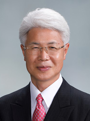
Takashi Akasaka
Nishinomiya Watanabe Cardiovascular Cerebral Center, Japan
Severe coronary calcification poses significant challenges in percutaneous coronary intervention (PCI), requiring advanced imaging and innovative procedural strategies to improve patient outcomes. At the Complex PCI 2024 conference, Takashi Akasaka, MD, delivered a comprehensive presentation detailing the latest techniques and tools for addressing this difficult subset of patients.
Challenges of Severe Coronary Calcification
Dr. Akasaka emphasized that severe coronary calcification significantly increases the risks of stent under-expansion, vessel damage, and restenosis. Lesions with circumferential calcium exceeding 180 degrees are particularly difficult to manage, necessitating a systematic approach involving imaging and calcium modification techniques.
The Role of Advanced Imaging
Intravascular imaging plays a crucial role in the successful treatment of calcified lesions. Optical coherence tomography (OCT) is particularly effective for visualizing calcium thickness and distribution, while intravascular ultrasound (IVUS) provides higher sensitivity in detecting calcium. Dr. Akasaka recommended a dual-modality approach combining OCT and IVUS for comprehensive lesion assessment and procedural planning.
Innovative Calcium Modification Techniques
Dr. Akasaka presented recent advances in lesion preparation, highlighting the importance of calcium modification techniques to facilitate optimal stent deployment. Tools such as rotational atherectomy, orbital atherectomy, intravascular lithotripsy (IVL), and scoring or cutting balloons were discussed as key options for addressing heavily calcified lesions.
IVL, in particular, was noted for its ability to fracture thick calcium layers exceeding 500 microns, which are often resistant to traditional balloon dilation. This technique has shown promising results in improving stent expansion and reducing the risk of restenosis.
Challenges with Calcified Nodules
Calcified nodules, particularly the "elapsed" type, remain a significant challenge in PCI. These lesions are associated with worse clinical outcomes and higher target lesion revascularization rates. Dr. Akasaka noted that while current treatment options such as drug-eluting stents (DES) and drug-coated balloons (DCB) have shown limited success, further innovation is required to address this unmet need.
Key Takeaways
Dr. Akasaka concluded the presentation with several take-home messages:
- Intravascular imaging with OCT and IVUS is essential for accurate assessment and treatment planning.
- Calcium modification techniques, such as IVL and atherectomy, are critical for procedural success in severely calcified lesions.
- The management of calcified nodules remains a pressing challenge, requiring continued research and innovation.
The insights shared at Complex PCI 2024 underscore the importance of combining advanced imaging with innovative procedural strategies to optimize outcomes in patients with severe coronary calcification. As the field continues to evolve, these advancements pave the way for better care and improved prognoses in this challenging patient population.
Live Case 3: Imaging and Physiology / Calcification
Friday, November 29, 12:40 PM ~ 2:30 PM
Main Arena
Edited by

Junghoon Lee, MD
The Catholic University of Korea, Eunpyeong St. Mary's Hospital, Korea (Republic of)

Takashi Akasaka
Nishinomiya Watanabe Cardiovascular Cerebral Center, Japan
Severe coronary calcification poses significant challenges in percutaneous coronary intervention (PCI), requiring advanced imaging and innovative procedural strategies to improve patient outcomes. At the Complex PCI 2024 conference, Takashi Akasaka, MD, delivered a comprehensive presentation detailing the latest techniques and tools for addressing this difficult subset of patients.
Challenges of Severe Coronary Calcification
Dr. Akasaka emphasized that severe coronary calcification significantly increases the risks of stent under-expansion, vessel damage, and restenosis. Lesions with circumferential calcium exceeding 180 degrees are particularly difficult to manage, necessitating a systematic approach involving imaging and calcium modification techniques.
The Role of Advanced Imaging
Intravascular imaging plays a crucial role in the successful treatment of calcified lesions. Optical coherence tomography (OCT) is particularly effective for visualizing calcium thickness and distribution, while intravascular ultrasound (IVUS) provides higher sensitivity in detecting calcium. Dr. Akasaka recommended a dual-modality approach combining OCT and IVUS for comprehensive lesion assessment and procedural planning.
Innovative Calcium Modification Techniques
Dr. Akasaka presented recent advances in lesion preparation, highlighting the importance of calcium modification techniques to facilitate optimal stent deployment. Tools such as rotational atherectomy, orbital atherectomy, intravascular lithotripsy (IVL), and scoring or cutting balloons were discussed as key options for addressing heavily calcified lesions.
IVL, in particular, was noted for its ability to fracture thick calcium layers exceeding 500 microns, which are often resistant to traditional balloon dilation. This technique has shown promising results in improving stent expansion and reducing the risk of restenosis.
Challenges with Calcified Nodules
Calcified nodules, particularly the "elapsed" type, remain a significant challenge in PCI. These lesions are associated with worse clinical outcomes and higher target lesion revascularization rates. Dr. Akasaka noted that while current treatment options such as drug-eluting stents (DES) and drug-coated balloons (DCB) have shown limited success, further innovation is required to address this unmet need.
Key Takeaways
Dr. Akasaka concluded the presentation with several take-home messages:
- Intravascular imaging with OCT and IVUS is essential for accurate assessment and treatment planning.
- Calcium modification techniques, such as IVL and atherectomy, are critical for procedural success in severely calcified lesions.
- The management of calcified nodules remains a pressing challenge, requiring continued research and innovation.
The insights shared at Complex PCI 2024 underscore the importance of combining advanced imaging with innovative procedural strategies to optimize outcomes in patients with severe coronary calcification. As the field continues to evolve, these advancements pave the way for better care and improved prognoses in this challenging patient population.
Live Case 3: Imaging and Physiology / Calcification
Friday, November 29, 12:40 PM ~ 2:30 PM
Main Arena
Edited by

Junghoon Lee, MD
The Catholic University of Korea, Eunpyeong St. Mary's Hospital, Korea (Republic of)

Leave a comment